Free shipping on orders over $500 throughout NSW and to all interstate Capitals.
Currency
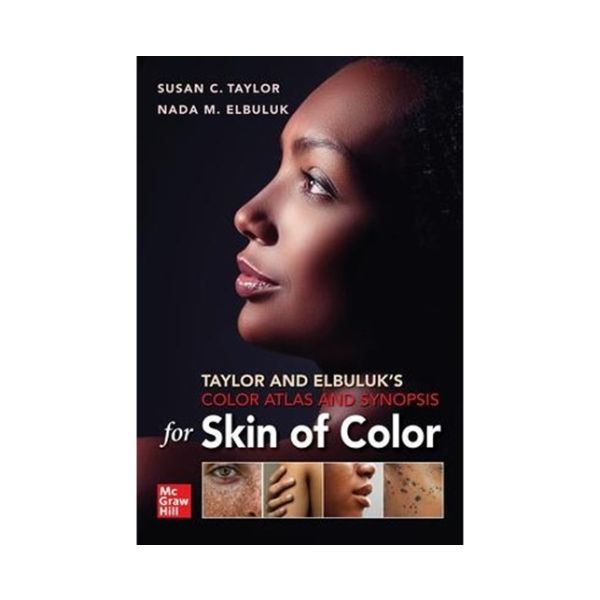
The expert guide to identifying and understanding the differences of common dermatology conditions in patients with all skin tones.
Dermatologists, medical students, and other health professionals untrained to recognize presentation of skin disease in non-white patients often inaccurately diagnose skin disease in skin of color patients. The perfect resource for comparative study of dermatologic disorders in skin of color, Taylor and Elbuluk's Color Atlas and Synopsis for Skin of Color helps readers recognize the differences in disease presentation amongst lighter and darker skin types. This understanding will ultimately lead to the provision of optimal care to patients with skin of color. Through hundreds of color images, this unique guide illustrates the differences in appearance of common dermatology conditions between Fitzpatrick’s Skin Type I-III light skin tones and Fitzpatrick’s Skin Type IV-VI dark skin tones.
| Author | Susan Taylor, MD and Nada Elbuluk, MD, MSc |
|---|---|
| Table Of Content | Inflammatory and Papulosquamous Disorders Atopic Dermatitis Morphological types Papular eczema Follicular accentuation Dyschromia Lichenification Pityriasis alba Psoriasis Morphology and color (violaceous and hyperpigmented with lichenification vs erythematous and devoid of silvery white scale) Contact Dermatitis Morpholology and color (hyperpigmentation vs erythema vs LPP-like in South Asian population) Pityriasis Lichenoides Chronica Morphology and color (hypopigmented patches or vitilgo like in darker skin vs erythematous guttate papular appearance in light skin) Pityriasis Rosea Morphology and color (similar distribution hyperpigmented to violeceous vs. erythematous; papular variant) Seborrheic Dermatitis a Facial Morphology and color (petaloid seborrheic dermatitis and hypopigmentation vs erythema) b. Scalp - Morphology and color (more scale vs sebopsoriasis like in lighter skin) Lichen Planus Morphology and color (similar morphology except hypertrophic LP darker violet or brown to black vs bright violet in lighter skin) Lichen nitidus (B) Color (Highlight differences in flesh color) Infections Tinea Versicolor Morphology and color (less pink or red or hypopigmented or hyperpigmented; sequelae of pigmentation remains) Tinea Capitis Morphology (kerion) Tinea Corporis Color (erythema vs Hyper- or hypopigmentation) Impetigo Morphology and color (erythema vs hyperpigmentation; both have honey colored crust) Cellulitis Color (lack of erythema) Syphilis Morphology (secondary syphilis with moth eaten alopecia; palmar lesions, facial rash) Verruca plana Color (skin colored papules that can be missed) Molluscum Color (skin colored papules that can be missed) 9a. COVID-19 Infestations/Bites Scabies Morphology and color (differences in location such as inter-digital; more erythematous) Pediculosis Erythema Migrans Color (lack of erythema vs hyperpigmentation and the intensity of violet hues) Drug Reactions DRESS Color (pigmentary differences) Morbilliform Drug Color (pigmentatio n difference both popular) Fixed Drug Color (lack of erythema vs hyperpigmented to black hue) Stevenâs Johnson Syndrome/Toxic Epidermal Necrolysis Morphology and color Follicular Disorders Acne Morphology and color (post-inflammatory hyperpigmentation and sequalae; definition of scarring) Rosacea Morphology and color (under-diagnosed in darker skin) Perioral dermatitis Morphology and color (pigmentation difference vs erythema Folliculitis Morphology and color (similar to acne) Hidradenitis Suppurativa Morphology and color (pigmentation difference vs erythema (keloidal scarring) Pseudofolliculitis Barbae Morphology and color (pigmentation difference vs erythema) Benign Neoplasms Seborrheic Keratosis and Dermatosis Papulosis Nigra Morphology and color (pink vs brown; size and distribution) Dermatofibroma Color (pink vs brown) Scars Morphology (hypertrophic scar vs keloid Malignancies Basal Cell Color (pigmented vs classical pink) Squamous Cell Color (pigmented vs classical pink) Melanoma Morphology and color (location acral and melanonychia) CTCL Morphology and color (hypopigmented Mycosis fungoides; follicular or sryingotrophic which may look like keratosis pilaris) Alopecias Lichen Plano Pilaris and Frontal Fibrosing Alopecia Morphology (triad of follicular papules, LPP and FFA in darker women) Traction Alopecia vs FFA Discoid Lupus Erythematosis Morphology and color (degree of hypopigmentation; follicular plugging) Folliculitis Decalvans Morphology and color (differences and keloid scarring Dissecting Cellulitis (B) Color differences Pigmentary Disorders Melasma Color (degree of pigmentation; telangectasias; confluence of pigmentation; extra-facial melasma) Postinflammatory Pigmentation vs PIE Photosensitivity PMLE(B) Chronicactinic dermatitis Morphology and color (more erythematous in white skin and darker and more lichenified in darker skin Vascular Disorders Purpura and vasculitis Morphology and color (violeceous) Miscellaneous Extrinsic Aging Morphology and color (rhytids and lentigines vs no lentigines and fine rhytids) Sarcoidosis Diabetes Mellitus Acanthosis Nigricans (color difference) Diabetic Dermopathy (color difference) Idiopathic Guttate Hypomelanosis Morphology and color (quantity and size in darker skin hues) Urticarial Bullous Pemphigoid Morphology and color |
| Format | Paperback |
| Page Count | 336 |